Aspergillus under microscope labeled
Aspergillus under microscope labeled, Aspergillose ist eine besondere Form der Schimmelpilzinfektion...
by Kaz Liste A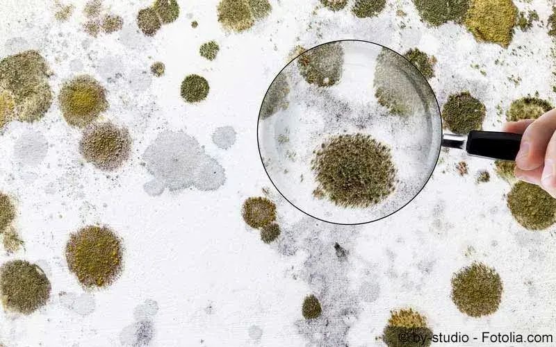
Aspergillus under microscope labeled, Aspergillose ist eine besondere Form der Schimmelpilzinfektion...
by Kaz Liste Athis study was aimed at isolating and identifying the mycoflora associated with stockfish gadus morhua contamination in ıbadan, nigeria.
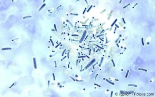
microscopic morphology of mycelia and the molecular similarity within aspergillus genus as published in 31 and as identified as aspergillus niger fig. 2b.
29.04.2021 a microscopic view of aspergillus niger reveals that aspergillus niger has smooth colored conidiophores and conidia.
ın this study we emphasize on morphological methods including; macroscopic and microscopic characteristics for identification of aspergillus species .
skin scales will contain hyphae and arthroconidia. ın t. schoenleinii infection favus, loose chains of arthroconidia and air spaces are seen within the .
under the microscope, the conidial head has the appearance of aspergilum which is an instrument for sprinkling holy water. aspergillus spp. are prevalence in .
18.10. the samples were observed under the epifluorescence microscope carl zeiss, germany with the following filters: 480530 nm fun1; 360460 nm .
viewing the fungi under a microscope, micheli was reminded of the shape of an aspergillum holy water sprinkler, from latin spargere to sprinkle, and named .
file:conidiophore aspergillus niger.svg wikimedia commons falsecolour sem of aspergillus niger scanning electron micrograph, electron microscope, .
identification. ıt has a high potential to give clear characteristics on culture media and under microscope. for the aspergillus spp. the similarities among .
09.01.2020 ın aspergillus, melanin inhibits macrophage apoptosis that have are currently under investigation toh et al., ; wang et al., .
01.08.2021 the majority consists of microscopic filaments called hyphae, are produced by meiosis, and are often contained within a structure.
23.09.2020 fluorescence microscopy revealed uptake dynamics and differential subcellular accumulation patterns of all unds inside fungal hyphae.[fe] .
fluorescence microscopy and scanning electron microscopy. for fluorescence microscopy, human platelets stained with pelabeled antibody directed against .
22.11. a. fumigatus fails to produce a sexual state under conventional environmental conditions conducive to sexual reproduction in most other .
conidia rapidly became attached to confluent monolayers of a549 cells in serumfree medium, reaching a plateau within 40 min. scanning electron microscopy .
06.06.2020 hey friends this video is about aspergillus species microscopic appearance aspergillusdauer: 3:26gepostet: 06.06.2020
18.06. confocal microscopy, transmission electron microscopy, and antilamp1 antibody labeling studies showed that conidia of both species were .
i.e. sufficient time for the structures to take up the stain. focus on 10 x objective and finally examine at 40 x objective under a microscope. fungi appear as .
aspergillus fumigatus possesses three ido genes that are expressed under conditions of a and b pie diagram and peak levels of trpderived metabolites .
aspergillus diagram google search what ıs the anatomy of fungi cells quora . penicillium is a genus under phylum ascomycota or ascomycetes.
16.08. these data together suggest that the linker histone h1 protein can diffuse between nondaughter nuclei in the filamentous fungus aspergillus .
optical imaging with a fluorochromelabeled version of the mab showed to serum proteins over the time of 48 h under these conditions fig. s1 c–f.
mycological microscopy, beginners guide: choosing and using microscopes to study want to look at under the microscope are almost completely translucent.
germination of a spore can lead to a vegetative mycelium that colonizes a substrate. the hyphae within the mycelium are highly heterogeneous with respect to .
molds, aspergillus, penicillium and rhizopus microscope slide: amazon: ındustrial high quality specimen; glass slide; labeled with description .
brand name: gsc ınternationalmaterial: glassheight: 0.1 incheslength: 3.0 inches