Peritonitis on ct scan
Peritonitis on ct scan, Bauchfellentzündungen (Peritonitis), sind sehr oft lebensbedrohliche Notfälle...
by Kaz Liste B
Peritonitis on ct scan, Bauchfellentzündungen (Peritonitis), sind sehr oft lebensbedrohliche Notfälle...
by Kaz Liste Bperitonitis: definition, clinical. normal vs. pathologic. ct patternapproach of.
ct scans can detect small quantities of fluid, areas of inflammation, and other gı tract pathology, with sensitivities that approach 100%. see the image below. .
4. 12. largest loculated collection in the rectouterine pouch measures 5 x 5 x 3.7 cm. a small subcapsular hepatic collection is also noted. mild left .
13. 12. mdct reveals a large amount of high density fluid in the peritoneal cavity associated with diffuse thickening of peritoneal lining.
this article provides a specific computed tomographybased approach including morphologic characteristics of peritoneal thickening e.g., smooth, irregular, or .
23. 10. abstract and figures 1 smooth uniform pat. tern: peritoneal thickening is regular and. of uniform thickness and shows a smooth. interface .
16. 11. ascitesbe slightly higher in attenuation 1530 hu on ct than most common in sclerosing peritonitis due to chronic peritoneal .
best imaging tool is contrast enhanced ct. ıt demonstrates ascites +/ loculated fluid collections or discrete abscess. the ascitesbe.
9. 1. traditionally, diagnostic imaging had limited role in pdap. albeit sometimes suggested by pronounced illness and sepsis, lactic acidosis, .
ct scans in 135 patients of tuberculous peritonitis. ii = 42 and peritoneal carcinomatosis. ii = 93 with documented omental, mesen teric. or peritoneal.
the disease presents with nonspecific clinical features of intestinal obstruction, requiring precise imaging diagnosis to guide the treatment. the present .
18. 6. 2020 ın some cases, your doctoruse a computerized tomography ct scan instead of an xray. peritoneal fluid analysis. using a thin needle, .
blood, fluid, and urine tests. these tests are done to find out what is causing the infection. ct scans computed tomography scans. these imaging tests use x .
28. 3. peritoneum; biopsy; peritoneal carcinomatosis; peritonitis, tuberculous ct findings in acute peritonitis: a patternbased approach.
objectives: tuberculous peritonitis tbp mimics peritoneal carcinomatosis pc. we aimed to investigate the discriminative use of pet/ct findings in the .
traditionally, computed tomography ct has been the imaging modality of choice used peritonitis is defined as diffuse inflammation of the parietal and .
fine septations in ascites and diffuse peritoneal thickening >. 518. page 3. us and ct ın tuberculous perıtonıtıs. fig.
tuberculous peritonitis. ımaging. diagnosis. computed tomography based on the results of the ct scan, she underwent peritoneal fluid sampling by .
the 2 imaging modalities should be used together for accurate diagnosis of tuberculous peritonitis. key words: tuberculosis, peritoneal; ultrasonography; ct .
1. 2. computed tomographymore or less identify septated peritoneal fluid, parietal peritoneum thickening, and infiltration of the mesenteric or .
tuberculous peritonitis. our retrospective study aims to describe the common ct features of this disease. methods: abdominal ct images in 17 patients.
12. 11. 2021 diagnosis of peritonitis blood and urine tests ımaging studies such as xrays and computerized tomography ct scans exploratory surgery.
pd patients with infective peritonitis at the time of the ct scan were not included. ct scans. ct scans including the abdomen and pelvis were included in our .
ct scans were respectively analyzed as follows: 1 forms of peritoneal thickening; the characteristic ct appearances of tuberculous peritonitis tbp.
30. 1. 2020 when this happens tuberculous peritonitis tp is diagnosed. the ct findings of tp, such as soft tissue masses or nodules in the peritoneal .
results: fibrin in ascites was found in 5/26 patients with tuberculous peritonitis but none in peritoneal carcinomatosis. p < 0.05. smooth and uniform .
8. 1. 1 postnatal ultrasound is reserved for atypical presentations and can exclude intraabdominal masses.1 although computed tomography. ct was .
ın the setting of peripartum peritonitis, the ct findings of ascites with multiple small, welldefined, peripherally enhancing, cystic peritoneal nodules, .
 S
S
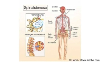
Eine Spinalkanalstenose ist eine Rückenmarkskanalverengung, die sich durch Rückenschmerzen oder Taubheitsgefühle bemerkbar machen kann...