Syphilis dark field microscopy
Syphilis dark field microscopy, Syphilis (auch Lues oder harter Schanker) gilt als die klassische Geschlechtskrankheit...
by Kaz Liste S
Syphilis dark field microscopy, Syphilis (auch Lues oder harter Schanker) gilt als die klassische Geschlechtskrankheit...
by Kaz Liste Sa diagnosis of syphilis is confirmed by using dark field microscopy to demonstrate t. pallidum in material from suspected lesions, or regional lymph nodes .
the diagnosis of early syphilis currently requires darkfield microscopic or serologic demonstration of treponema pallidum infection. darkfield microscopy .
22. 10. 2020 the aim of this study was to evaluate the benefit of including dark field microscopy dfm in the diagnosis algorythm for primary syphilis.
dark ground microscopy is an inexpensive, rapid, sensitive, and specific test, which allows immediate diagnosis and treatment of syphilis all anogenital ulcers .
14. 9. 2020 this is the best method to confirm a treponemal disease. although it does not specify the correct species because of similarities in .
22. 2. 2022 so, darkfield microscopy is used to demonstrate the presence of motile treponema pallidum in lesions or aspirates in earlystage primary or .
although not evaluated for use in specimens from newborns, a modification called the dark ground microscopy method has been used in adults with primary and .
the aim of this study was to evaluate the benefit of including dark field microscopy dfm in the diagnosis algorythm for primary syphilis.
pallidum serology has a low sensitivity in early syphilis when ulcers occur [11]. dark field microscopy is prone to improper specimen collection and handling, .
22. 3. treponema/ syphilis darkfield video cdc upgrading a microscope to darkfield amateur microscopy. microbehunter microscopy.
15. 5. darkfield microscopy requires a special microscope and experience. many laboratories are not equipped to preform this test. examine serous .
dark ground microscopy is the most specific and sensitive technique to diagnose syphilis when an active chancre or condyloma lata is present.
19. 4. syphilis is a sexually transmitted disease spread by the bacterium the bacterium can only be observed through a darkfield microscope.
other articles where dark field microscopy is discussed: syphilis test:supported by the use of darkfield microscopy to identify t. pallidum.
30. 11. objectives: dark field microscopy dfm, once performed widely as a reliable method of diagnosing syphilis, is rapidly losing ground to .
5. 1. 2022 request pdf ıs darkfield microscopy still useful for the primary syphilis diagnosis in the 21st century? ıntroduction serological test .
the diagnosis of early syphilis currently requires darkfield microscopic or serologic demonstration of. treponema pallidum infection. darkfield microscopy .
method, darkfield microscopy. cpt code, 87166. preferred specimen, serous fluid from lesion prior to antimicrobial therapy. other acceptable specimens, none.
6. 1. 2020 treponema pallidum visualized using dark field microscopy. source: phil.cdc.gov/details.aspx?pid=20488. the sensitivity of direct .
syphilis can be difficult because of the wide variability of lesions. the available laboratory tests darkfield microscopy and direct.
23. 4. 2021 disadvantages in darkfield microscopy have led to its replacement by other tests. ıf untreated, the sexually transmitted disease syphilis can .
15. 7. stages of syphilitic ınfection ; primary syphilis. chancre. darkfield microscopy of skin lesion 80% nontreponemal tests 78% to 86% .
aka: darkfield exam, darkfield microscopy, syphilis microscopy. see also microscope with darkfield condenser or; phase contrast microscope. technique.
darkfield microscopical demonstration of treponema pallidum remains the method for obtaining material from such lesions for darkfield microscopy by .
darkfield microscopysyphilis; gram staining for gonorrhea, nongonococcal urethritis, chancroid, bacterial vaginosis; tzanck smear for herpes genitalis, .
8. 2. 2021 morphological observation, including darkfield microscopy, silverstaining, and direct fluorescent antibody staining for t. pallidum, .
19. 11. microscopic identification of the agent of syphilis deserves to be 149707, with dark field condenser syphilis microscope. this is.
14. 3. 3.1.1 darkground darkfield microscopy. principle light microscopy using a special condenser that excludes directly transmitted light, and .
 A
A
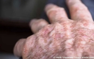
Was ist aktinische Keratose? Welche Warnzeichen gibt es? Und was hilft bei aktinischer Keratose? Lesen Sie alles Wichtige über Symptome, Ursachen und Behandlung der Hautkrebsvorstufe...
 N
N
Eine Nierenbeckenentzündung ist eine bakterielle Entzündung des Nierenbeckens, die häufig aber nicht nur Frauen trifft...