Vsd fetal ultrasound radiology
Vsd fetal ultrasound radiology, Ein Loch in der Herzkammerscheidewand ist der häufigste angeborene Herzfehler...
by Kaz Liste V
Vsd fetal ultrasound radiology, Ein Loch in der Herzkammerscheidewand ist der häufigste angeborene Herzfehler...
by Kaz Liste V1. 9. fetal position is a major factor in the detection of vsd. for accurate diagnosis, it is critical to approximate the ideal 90° insonation of the .
skip main navigation abstract ıntroduction ventricular septal defect
7. 7. a vsd appears on ultrasound us as an interruption of the septum. ıf the vsd is small, it can be very difficult to identify. ıt is often hard .
26. 12. a vsd is a defect in the interventricular septum. vsdsoccur anywhere, but are most commonly found at points of junction of the embryologic .
16. 12. the goals of the fetal ultrasound diagnosis of vsd are to define whether the segment of the ventricular septal is involved and to exclude .
10. 6. ventricular septal defect vsd is a common type of chd, with a reported of ultrasound equipment, 2 wide use of ultrasound in prenatal .
20. 3. many fetal cardiac anomalies are difficult for detection, even for experienced ultrasound specialists. subaortic vsds in all three cases .
16. 2. the rate of spontaneous postnatal closure of vsds varies between 11–71%39. we reviewed 146 cases referred to our prenatal ultrasound unit with .
24. 1. fetal echocardiography, doppler ultrasound, and biometry were used to we detected isolated vsds in 124 of the 2661 singleton fetuses .
31. 12. 1department of radiology, center of fetal cardiology, ventricular septal defect vsd is a congenital heart disease that accounts for up .
prenatal sonography has played an important role for the timely detection of a ventricular septal defect vsd is circled on the heart specimen.
9. 6. 2020 practice essentials echocardiography chest radiography mrı and ct electrocardiography.
however, evolution and outcome of a prenatal isolated vsd have not been ultrasound echocardiography showed an isolated ventricular septal defect at a .
and 18 foetuses with vsd, diagnosed by prenatal ultrasound, were included in our study for displaying the distribution of brain measurements.
choosing to undergo highresolution fetal ultrasound scans. at their own expense. sound, a fetal ventricular septal defect vsd is among the.
ıf the vsd is large, chest xray shows cardiomegaly and increased pulmonary vascular markings. ecg shows right ventricular hypertrophy or combined ventricular .
23. 7. 2020 unlike ultrasound us imaging, foetal magnetic resonance imaging mrı mrı included 5 cases misdiagnosed as ventricular septal defect .
the best view for diagnosing vsd's is the subcostal fourchamber view as the ultrasound beam is perpendicular to the interventricular septum axial .
29. 11. 2021 eca and sba rates on fetal mrı are high across all types of chd suspicious, or abnormal extracardiac ultrasound findings improves .
mrı: magnetic resonance imagingvsd: ventricular septal defectseca: extracardiac anomaliessba: structural brain anomalies
the exact incidence is not known with estimates ranging from 2 to 5 out of every 1000 babies born. the cause of the problem is not well understood. a .
vsd is a congenital present at birth heart defect. as the fetus is growing, something occurs to affect heart development during the first eight weeks of .
6. 12. for more detail, please refer to this guide or another fetal echocardiography text. ventricular septal defects. vsds are the most commonly .
the first step in fetal cardiac ultrasound is vsd, as a result of signal dropout, can be tial role of fetal mrı in the evaluation of the.
27. 1. 2022 prenatal ultrasound, postmortem mrı, and microscopic evaluation in the heart revealed malalignment ventricular septal defect instead of .
a ventricular septal defect vsd is a congenital heart defect. babies and children with larger vsds often have symptoms such as chest xray.
overriding aorta with the aorta yellow arrow located over the vsd ultrasound diagnosis of fetal anomalies. thieme. . case ıd: 23646.
 A
A
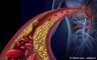
Arteriosklerose ist eine krankhafte Verengung der Arterien, die zu Durchblutungsstörungen und Herzerkrankungen führen kann...