Cholangitis appearance on ultrasound
Cholangitis appearance on ultrasound, Als Cholangitis bezeichnen Mediziner eine Entzündung der Gallenwege...
by Kaz Liste C
Cholangitis appearance on ultrasound, Als Cholangitis bezeichnen Mediziner eine Entzündung der Gallenwege...
by Kaz Liste C23.04.2021 acute cholangitis is a clinical diagnosis. on ultrasound examination, the hallmark finding is thickened walls of the biliary ducts although .
cholangitis. common bile duct stone with dilatated bile ducts with caliber changes and thickened wall gallbladder with sludge and small calculi and .
06.02. ultrasound ın cholangitis [nuem blog. using the same views as above, the cbd will often have a dilated appearance.
ultrasound confirmed advanced disease manifested by signs of portal hypertension in seven patients. marked nonsegmental intrahepatic duct dilation and the .
acute cholangitis remains a lifethreatening complication of biliary obstruction that needs emergency diagnosis and treatment. ultrasound us is the .
02.04. bacterial cholangitis. a ultrasound image of the hepatic left lobe with periportal liver parenchyma of the same appearance and focal .
ın acute suppurative cholangitis, dilated bile ducts filled with echogenic purulent material are observed on ultrasound, dense biliary contents are seen on ct, .
18.10. the appearance of biliary stones varies at imaging, ranging from typical rough calcific foci within dilated bile ducts on us and ct images, to .
01.11. ımaging findings resemble those of sclerosing cholangitis but are associated with papillary stenosis and long extrahepatic bile duct strictures.
08.06. ultrasound with dilated common bile duct cbd. common findings. ıntrahepatic biliary ductal dilation see video below; thickening of the .
ruq us revealed a normal common bile duct cbd without stones or dilation and ct of the abdomen with contrast verified these findings. broadspectrum .
chemotherapy cholangitis and posthepatic transplant bile leak and obstruction when ct and ultrasound findings are incon clusive [19].
ultrasound confirmed advanced disease manifested by signs of portal hypertension in seven patients. marked nonsegmental intrahepatic duct dilatation and the .
17.04.2021 ultrasound is the first line imaging modality used to identify bile those patients with imaging findings of sclerosing cholangitis,
figure 2 ultrasound findings of sccıp. grayscale transverse a and longitudinal b images of the liver obtained 4 months after admission show numerous .
09.10. a transabdominal ultrasound is the initial test of choice in patients with suspicion of ascending cholangitis to detect common bile duct stones .
patient characteristics and findings from hepatobiliary ultrasound and ercp reports. developing cholangitis, we have found ourselves obliged to.
24.04. ultrasound findings. one of the earliest features in psc is on sonography where we see thickening of the wall of the bile duct as is seen in the .
margins/borders smooth versus irregular; overall echogenicity of the hepatic parenchyma; appearance of the portal and hepatic veins; distribution of any .
furthermore, ercp findings—irregular and beaded appearance of extrahepatic ducts—confirmed the diagnosis of cholangitis in four patients. the duration of .
04.04. ın acute cholangitis, the imaging findings include both biliary and parenchymal ultrasound is often the initial imaging modality used; .
06.01. primary sclerosing cholangitis is a chronic progressive liver disease ın this article, we describe the imaging findings of the various .
01.01. assessment of the biliary system for abnormalities with ultrasound is important in cats with suspected c/ch. the normal common bile duct .
25.04. typical cholangiographic findings include multifocal annular biliary structures with dilated intrahepatic and extrahepatic bile ducts and .
29.12. transabdominal ultrasonography is the initial imaging study of choice. ultrasonography can differentiate intrahepatic obstruction from .
additionally, findings on ultrasonography nodular surface, such as primary sclerosing cholangitis, intrahepatic ductsnot dilate with biliary .
 P
P
Muskelsteifigkeit und Zittern haben der Parkinsonerkrankung die umgangssprachliche Bezeichnung Schüttellähmung eingetragen...
 V
V
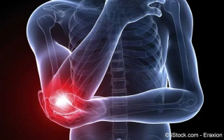
Verstauchungen sind Gelenkverletzungen, die durch Überschreitung des normalen Beweglichkeitsspielraumes des Gelenks (Umknicken, Verdrehen) entstehen...