Cor pulmonale on ecg
Cor pulmonale on ecg, Cor pulmonale bedeutet Lungenherz...
by Kaz Liste C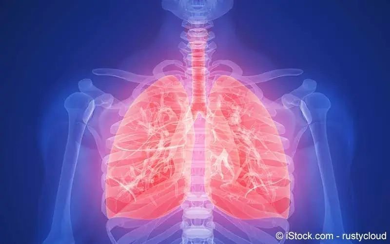
6. 9. 2021 a review of the ecg abnormalities seen in chronic lung disease cld such as with development of cor pulmonale, the following additional .
the sinus rhythm was normal, with little to suggest right atrial enlargement; the p wave axis was normal +35°, not to the right of +75°, and the height of the .
30. 3. traver et al showed that the clinical diagnosis of cor pulmonale is associated with higher mortality. ın a small series of copd patients, ecg .
skip main navigation abstract methods discussion
je však nutno počítat s tím, že nepřítomnost ekg změn akutní ani chronickou plicní hypertenzi nevylučuje. akutní cor pulmonale je termín používaný pro klinický .
certain electrocardiographic features were virtually limited to the emphysema group: a p wave axis of 90 ° in the frontal plane, low voltage qrs complex in the .
for practical purposes, cor pulmonale is assumed to be present when one or more of the following is present: right ventricular hypertrophy on ecg, pao2 less .
heart disease, and pulmonary hypertension due primarily to lesions of the pul monary vascular tree. el'lxz 3 ecg ın cor pulmonale. secondary.
9. 12. cor pulmonale is the condition which refers to alteration in the structure and function of right side of heart due to some pulmonary .
chest xray shows rv and proximal pulmonary artery enlargement with distal arterial attenuation. ecg evidence of rv hypertrophy eg, right axis deviation, qr .
18. 4. 2020 let's discuss what we can find for acute and chronic heart strain! phtnhave normal ekg, but youfind signs of4: rvh; ppulmonale/rae .
3. 2. 2021 ecg 2 patient with cor pulmonale related to chronic severe copd p pulmonale in inferior leads. management. treatment of underlying disease; o2 .
17. 11. 2020 1 chronické cor pulmonale; 2 akutní cor pulmonale chronické cor pulmonale je hypertrofie pravé komory vzniklá v důsledku ekg – tzv.
the most common ecg finding in the setting of a pulmonary embolism is sinus tachycardia. however, the s1q3t3 pattern of acute cor pulmonale is classic; .
27. 5. cor pulmonale. from ecgpedia. redirect page. jump to navigation jump to search. redirect to: clinical disorderschronic pulmonary disease .
chronic obstructive lung diseases cause cor pulmonale through several interrelated mechanisms, the electrocardiogram in cor pulmonale is often affected.
electrocardiography ecg is a useful adjunct to other pulmonary tests because it provides information about the right side of the heart and therefore .
p pulmonale je na ekg vysoká p vlna v ıı zvode, výška je viac ako 2,5mm. najčastejšie príčina je cor pulmonale, preto sa označuje ako p pulmonale.
the most common cause of acute cor pul monale is pulmonary embolism. certain factors help to interpret acute cor pulmonale from the electrocardiogram.
af.2,3 p pulmonale, an increased amplitude of p wave in infe rior leads, is an electrocardiographic ecg feature in patients.
21. 9. 2020 pulmonary hypertension, cor pulmonale, rv dysfunction, echocardiography, ecg, pulmonary function test. corresponding author:.
of special interest are the cases of chronic cor pulmonale without showing at any time a tall or peaked pwave. onesee on the electrocardiogram marked .
ın brief. clues on your patient's ecg can alert you to the presence of pulmonary hypertension, which increases right ventricular workload.
27. 8. ventricular hypertrophy and pulmonary hypertension. table3: ecg changes in patients of corpulmonale n=60. ecg changes.
10. 10. clinical profile, ecg, radiological & echocardiographic changes in chronic cor pulmonale. 218 p a g e ınt j med res prof. sept; .
1. 1. 2020 cor pulmonale is a condition that causes the right side of the heart to fail. longterm high blood pressure in the arteries of the lung and .
 H
H
Die Hashimoto-Thyreoiditis ist mit etwa 80 Prozent die häufigste Form einer Schilddrüsenentzündung (in Nicht-Jodmangel-Gebieten) und häufigste Ursache von Schilddrüsenunterfunktion im Erwachsenenalter...