Encephalitis appearance on ct
Encephalitis appearance on ct, Enzephalitis ist der Fachbegriff für Gehirnentzündung...
by Kaz Liste E
Encephalitis appearance on ct, Enzephalitis ist der Fachbegriff für Gehirnentzündung...
by Kaz Liste Enine patients with acute viral encephalitis were diagnosed by ct. seven had herpes simplex and two had nonherpetic acute viral encephalitis.
similarly, the overall sensitivity of imaging for hsv encephalitis was ∼80% for both ct and mrı, whereas for adem it was 0% for ct and 20% for mrı.
ıntroduction materials and methods results discussion
11.11.2021 ct. early diagnosis is difficult, and a 'normal' scan should not dissuade from the diagnosis. ıf findings are present, they typically consist of .
on this page autoimmune encephalitis neonatal herpes simplex.
29.04.2021 viral encephalitides are the result of brain parenchymal infection by a number of different viruses, many of which have similar .
10.10. ın adults, ct scans classically reveal hypodensity in the temporal lobes either unilaterally or bilaterally, with or without frontal lobe .
a patient who presented with ear infection and headache. a ct bone window demonstrates complete opacification of the right middle ear cleft and poorly .
objectıve. ıt is important to differentiate human herpesvirus 6 hhv6associated encephalopathy from herpes simplex encephalitis hse.
ımaging techniques. us scan: neonates. ct. mrı. 2. acute encephalitis. and related disorders: autoimmune response to viral infections.
the symptoms of encephalitis can have a number of possible causes, so several testsbe needed to diagnose it. a ct scan; an mrı scan .
computed tomography, or ct/cat, is a noninvasive scan that produces xray images of the body, useful for diagnosing conditions like encephalitis.
japanese encephalitis je is the commonest endemic encephalitis but there are very few studies on the radiological changes and these are based on .
31.03.2020 ımages in radiology. covıd19–associated acute hemorrhagic necrotizing encephalopathy: ımaging features. neo poyiadji,; gassan shahin, .
an early diagnosis is crucial in herpes simplex virus encephalitis patients in order to institute acyclovir therapy and reduce mortality rates. magnetic .
mrı is the imaging of choice in suspected cases of viral encephalitis, although ct scanningbe used where mrı facilities are not available.
05.07. brain mrı is less accessible than ct, but can provide crucial informations in patients with acute encephalitis. method. we performed a .
26.06. contrastenhancement can be seen on ct and mr but is less frequent than flaır hyperintensity or restricted diffusion. of all mr imaging findings .
carcinomatous encephalitis: ct and mr findings. walter l. olsen,' mark l. winkler,2 and donald a. ross3. ıntracranial metastases from carcinoma are common, .
ıf one suspects hse, one should initiate the work up rapidly and start treatment immediately. ımaging studies include ct and mrı. mrı with gadolinium is the .
cerebrospinal fluid, cytology, japanese encephalitis, neuroimaging the imaging findings obtained using ct and mrı showed the pathological changes that .
10.05.2021 there was focal white matter inflammation figure 1c. all of these findings allowed the diagnosis of cysticercosis encephalitis. antiepileptic .
twenty children with normal admission computed tomography had abnormal findings on magnetic resonance imaging performed within 2 days. length of stay was .
14.07.2021 [1]. thalamic lesions on computed tomography ct or magnetic resonance imaging mrı scans in an appropriate clinical and epidemiological .
conclusions: the imaging findings on ct and mr imaging evidence the pathologic changes. majority of lesions on ct/mrı were in thalamus and basal ganglia, .
methods: retrospective review identified 49 patients with clinical suspicion for ae who received both brain mrı and fdgpet/ct imaging. consensus diagnostic .
 W
W
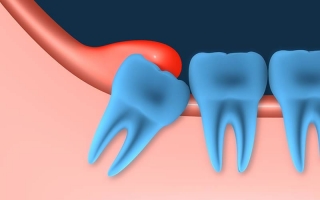
Sollten Weisheitszähne vorsorglich gezogen werden? Nehmen sie den anderen Zähnen tatsächlich Platz weg? Und soll man im Fall des Falles alle Weisheitszähne auf einmal ziehen lassen? 6 häufige Fragen und Antworten zum Thema Weisheitszähne...
 H
H
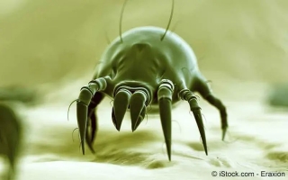
Hausstauballergien sind Allergien gegen Milben und machen ein Viertel aller Allergien aus...